

What Type Of Contraction Involves The Development Of Tension But No Change In Length

Hierarchical organization of skeletal musculus

Contractions of skeletal muscles permit vertebrate animals such as frogs to move
Muscle contractions underlie motion
Muscle wrinkle is the activation of tension-generating sites within musculus cells.[1] [2] In physiology, muscle wrinkle does non necessarily mean musculus shortening because muscle tension can be produced without changes in muscle length, such every bit when holding a heavy book or a dumbbell at the same position.[1] The termination of muscle contraction is followed by muscle relaxation, which is a return of the muscle fibers to their low tension-generating state.[i]
Muscle contractions can exist described based on two variables: length and tension.[1] A musculus contraction is described as isometric if the musculus tension changes just the musculus length remains the same.[ane] [3] [4] [5] In dissimilarity, a muscle wrinkle is isotonic if muscle tension remains the same throughout the contraction.[1] [iii] [4] [v] If the muscle length shortens, the contraction is concentric;[one] [half dozen] if the muscle length lengthens, the contraction is eccentric. In natural movements that underlie locomotor action, musculus contractions are multifaceted as they are able to produce changes in length and tension in a time-varying manner.[vii] Therefore, neither length nor tension is probable to remain the same in muscles that contract during locomotor activity.
In vertebrates, skeletal muscle contractions are neurogenic as they require synaptic input from motor neurons. A single motor neuron is able to innervate multiple muscle fibers, thereby causing the fibers to contract at the same fourth dimension. Once innervated, the protein filaments within each skeletal musculus fiber slide by each other to produce a wrinkle, which is explained past the sliding filament theory. The contraction produced tin can be described as a twitch, summation, or tetanus, depending on the frequency of action potentials. In skeletal muscles, muscle tension is at its greatest when the musculus is stretched to an intermediate length every bit described by the length-tension relationship.
Different skeletal musculus, the contractions of smooth and cardiac muscles are myogenic (meaning that they are initiated by the smoothen or centre muscle cells themselves instead of being stimulated by an exterior consequence such every bit nerve stimulation), although they can be modulated by stimuli from the autonomic nervous system. The mechanisms of contraction in these muscle tissues are similar to those in skeletal muscle tissues.
Types [edit]

Types of musculus contractions
Muscle contractions tin exist described based on two variables: force and length. Force itself can be differentiated as either tension or load. Musculus tension is the force exerted by the muscle on an object whereas a load is the force exerted by an object on the musculus.[1] When muscle tension changes without any corresponding changes in muscle length, the muscle contraction is described as isometric.[1] [3] [4] [5] If the musculus length changes while muscle tension remains the same, and so the muscle contraction is isotonic.[1] [3] [4] [v] In an isotonic contraction, the musculus length can either shorten to produce a concentric contraction or lengthen to produce an eccentric wrinkle.[1] [half-dozen] In natural movements that underlie locomotor activity, muscle contractions are multifaceted every bit they are able to produce changes in length and tension in a fourth dimension-varying manner.[vii] Therefore, neither length nor tension is likely to remain constant when the musculus is active during locomotor activeness.
Isometric wrinkle [edit]
An isometric contraction of a muscle generates tension without irresolute length.[1] [3] [4] [five] An instance tin be plant when the muscles of the hand and forearm grip an object; the joints of the manus exercise not movement, but muscles generate sufficient force to prevent the object from being dropped.
Isotonic contraction [edit]
In isotonic contraction, the tension in the muscle remains abiding despite a change in muscle length.[i] [3] [4] [5] This occurs when a muscle's force of contraction matches the total load on the muscle.
Concentric contraction [edit]
In concentric contraction, muscle tension is sufficient to overcome the load, and the muscle shortens as it contracts.[viii] This occurs when the strength generated by the muscle exceeds the load opposing its wrinkle.
During a concentric contraction, a muscle is stimulated to contract according to the sliding filament theory. This occurs throughout the length of the muscle, generating a strength at the origin and insertion, causing the muscle to shorten and changing the angle of the joint. In relation to the elbow, a concentric wrinkle of the biceps would cause the arm to bend at the elbow every bit the hand moved from the leg to the shoulder (a biceps roll). A concentric contraction of the triceps would change the angle of the joint in the opposite direction, straightening the arm and moving the hand towards the leg.
Eccentric wrinkle [edit]
In eccentric contraction, the tension generated while isometric is insufficient to overcome the external load on the muscle and the muscle fibers lengthen as they contract.[9] Rather than working to pull a joint in the direction of the muscle contraction, the muscle acts to decelerate the articulation at the end of a motion or otherwise control the repositioning of a load. This can occur involuntarily (e.g., when attempting to move a weight as well heavy for the muscle to lift) or voluntarily (east.one thousand., when the muscle is 'smoothing out' a movement or resisting gravity such as during downhill walking). Over the short-term, force training involving both eccentric and concentric contractions announced to increment muscular forcefulness more than than grooming with concentric contractions lonely.[10] However, exercise-induced muscle harm is also greater during lengthening contractions.[11]
During an eccentric contraction of the biceps musculus, the elbow starts the movement while aptitude and then straightens as the hand moves away from the shoulder. During an eccentric contraction of the triceps muscle, the elbow starts the movement straight and so bends every bit the hand moves towards the shoulder. Desmin, titin, and other z-line proteins are involved in eccentric contractions, but their machinery is poorly understood in comparison to crossbridge cycling in concentric contractions.[9]
Though the musculus is doing a negative amount of mechanical piece of work, (piece of work is existence done on the muscle), chemical energy (originally of oxygen,[12] unlocked by fat or glucose, and temporarily stored in ATP) is nevertheless consumed, although less than would be consumed during a concentric contraction of the same force. For example, one expends more than energy going up a flying of stairs than going down the same flying.
Muscles undergoing heavy eccentric loading suffer greater harm when overloaded (such as during muscle building or forcefulness training exercise) as compared to concentric loading. When eccentric contractions are used in weight training, they are ordinarily chosen negatives. During a concentric wrinkle, muscle myofilaments slide past each other, pulling the Z-lines together. During an eccentric contraction, the myofilaments slide past each other the contrary way, though the bodily move of the myosin heads during an eccentric contraction is not known. Exercise featuring a heavy eccentric load can actually back up a greater weight (muscles are approximately xl% stronger during eccentric contractions than during concentric contractions) and too results in greater muscular damage and delayed onset muscle soreness one to two days after training. Exercise that incorporates both eccentric and concentric muscular contractions (i.e., involving a strong wrinkle and a controlled lowering of the weight) can produce greater gains in strength than concentric contractions alone.[ten] [xiii] While unaccustomed heavy eccentric contractions can hands pb to overtraining, moderate preparation may confer protection against injury.[10]
Eccentric contractions in motility [edit]
Eccentric contractions unremarkably occur equally a braking force in opposition to a concentric contraction to protect joints from damage. During virtually any routine movement, eccentric contractions assist in keeping motions smooth, but tin also slow rapid movements such as a dial or throw. Function of training for rapid movements such every bit pitching during baseball involves reducing eccentric braking allowing a greater power to be adult throughout the movement.
Eccentric contractions are beingness researched for their ability to speed rehabilitation of weak or injured tendons. Achilles tendinitis[14] [xv] and patellar tendonitis[16] (likewise known as jumper'due south knee or patellar tendonosis) have been shown to benefit from high-load eccentric contractions.
Vertebrates [edit]

In vertebrate animals, in that location are 3 types of muscle tissues: 1) skeletal, ii) polish, and iii) cardiac
In vertebrate animals, there are three types of muscle tissues: skeletal, smooth, and cardiac. Skeletal muscle constitutes the bulk of musculus mass in the body and is responsible for locomotor activity. Smoothen muscle forms blood vessels, alimentary canal, and other areas in the body that produce sustained contractions. Cardiac muscle make up the middle, which pumps blood. Skeletal and cardiac muscles are called striated muscle because of their striped appearance under a microscope, which is due to the highly organized alternate blueprint of A bands and I bands.
Skeletal musculus [edit]

Organisation of skeletal muscle
Excluding reflexes, all skeletal muscles contractions occur equally a result of signals originating in the brain. The brain sends electrochemical signals through the nervous organization to the motor neuron that innervates several muscle fibers.[17] In the case of some reflexes, the signal to contract can originate in the spinal cord through a feedback loop with the grey matter. Other actions such equally locomotion, animate, and chewing accept a reflex aspect to them: the contractions tin be initiated both consciously or unconsciously.
Neuromuscular junction [edit]

Structure of neuromuscular junction.
A neuromuscular junction is a chemic synapse formed by the contact between a motor neuron and a muscle fiber.[18] It is the site in which a motor neuron transmits a signal to a muscle fiber to initiate muscle wrinkle. The sequence of events that results in the depolarization of the muscle fiber at the neuromuscular junction begins when an action potential is initiated in the cell body of a motor neuron, which is then propagated past saltatory conduction along its axon toward the neuromuscular junction. Once it reaches the terminal bouton, the action potential causes a Ca 2+
ion influx into the terminal by way of the voltage-gated calcium channels. The Ca 2+
influx causes synaptic vesicles containing the neurotransmitter acetylcholine to fuse with the plasma membrane, releasing acetylcholine into the synaptic cleft between the motor neuron terminal and the neuromuscular junction of the skeletal muscle fiber. Acetylcholine diffuses across the synapse and binds to and activates nicotinic acetylcholine receptors on the neuromuscular junction. Activation of the nicotinic receptor opens its intrinsic sodium/potassium channel, causing sodium to blitz in and potassium to trickle out. As a event, the sarcolemma reverses polarity and its voltage quickly jumps from the resting membrane potential of -90mV to equally high as +75mV as sodium enters. The membrane potential and then becomes hyperpolarized when potassium exits and is then adjusted dorsum to the resting membrane potential. This rapid fluctuation is chosen the terminate-plate potential[19] The voltage-gated ion channels of the sarcolemma next to the end plate open in response to the finish plate potential. They are sodium and potassium specific and just allow one through. This wave of ion movements creates the activeness potential that spreads from the motor end plate in all directions.[19] If action potentials stop arriving, and then acetylcholine ceases to be released from the terminal bouton. The remaining acetylcholine in the synaptic crevice is either degraded past active acetylcholine esterase or reabsorbed past the synaptic knob and none is left to supplant the degraded acetylcholine.
Excitation–contraction coupling [edit]
Excitation–contraction coupling is the process by which a muscular action potential in the muscle fiber causes the myofibrils to contract.[20] In skeletal musculus, excitation–wrinkle coupling relies on a directly coupling between key proteins, the sarcoplasmic reticulum (SR) calcium release channel (identified equally the ryanodine receptor ane, RYR1) and voltage-gated L-type calcium channels (identified as dihydropyridine receptors, DHPRs). DHPRs are located on the sarcolemma (which includes the surface sarcolemma and the transverse tubules), while the RyRs reside across the SR membrane. The close apposition of a transverse tubule and ii SR regions containing RyRs is described as a triad and is predominantly where excitation–contraction coupling takes place. Excitation–wrinkle coupling occurs when depolarization of skeletal musculus prison cell results in a musculus action potential, which spreads across the jail cell surface and into the muscle fiber's network of T-tubules, thereby depolarizing the inner portion of the muscle fiber. Depolarization of the inner portions activates dihydropyridine receptors in the terminal cisternae, which are in close proximity to ryanodine receptors in the adjacent sarcoplasmic reticulum. The activated dihydropyridine receptors physically collaborate with ryanodine receptors to actuate them via foot processes (involving conformational changes that allosterically activates the ryanodine receptors). As the ryanodine receptors open, Ca 2+
is released from the sarcoplasmic reticulum into the local junctional space and diffuses into the bulk cytoplasm to cause a calcium spark. Note that the sarcoplasmic reticulum has a big calcium buffering capacity partially due to a calcium-bounden protein called calsequestrin. The nearly synchronous activation of thousands of calcium sparks past the activity potential causes a cell-broad increase in calcium giving rise to the upstroke of the calcium transient. The Ca 2+
released into the cytosol binds to Troponin C by the actin filaments, to let crossbridge cycling, producing force and, in some situations, motion. The sarco/endoplasmic reticulum calcium-ATPase (SERCA) actively pumps Ca two+
back into the sarcoplasmic reticulum. As Ca 2+
declines back to resting levels, the force declines and relaxation occurs.
Sliding filament theory [edit]
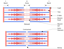
Sliding filament theory: A sarcomere in relaxed (above) and contracted (beneath) positions
The sliding filament theory describes a process used by muscles to contract. It is a cycle of repetitive events that cause a thin filament to slide over a thick filament and generate tension in the musculus.[21] It was independently developed by Andrew Huxley and Rolf Niedergerke and by Hugh Huxley and Jean Hanson in 1954.[22] [23] Physiologically, this wrinkle is not uniform beyond the sarcomere; the primal position of the thick filaments becomes unstable and can shift during contraction. However the actions of elastic proteins such every bit titin are hypothesised to maintain compatible tension across the sarcomere and pull the thick filament into a cardinal position.[24]
Cantankerous-bridge cycle [edit]
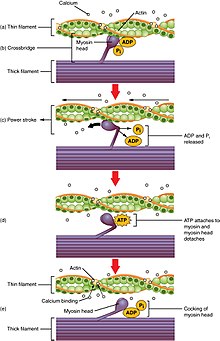
Cantankerous-bridge cycling is a sequence of molecular events that underlies the sliding filament theory. A cantankerous-bridge is a myosin projection, consisting of two myosin heads, that extends from the thick filaments.[1] Each myosin head has ii bounden sites: one for ATP and another for actin. The binding of ATP to a myosin head detaches myosin from actin, thereby allowing myosin to demark to some other actin molecule. Once attached, the ATP is hydrolyzed by myosin, which uses the released energy to movement into the "artsy position" whereby information technology binds weakly to a part of the actin bounden site. The remainder of the actin binding site is blocked by tropomyosin.[25] With the ATP hydrolyzed, the artsy myosin head at present contains ADP + Pi. Two Ca 2+
ions bind to troponin C on the actin filaments. The troponin-Ca 2+
complex causes tropomyosin to slide over and unblock the remainder of the actin binding site. Unblocking the balance of the actin binding sites allows the two myosin heads to close and myosin to bind strongly to actin.[25] The myosin head then releases the inorganic phosphate and initiates a power stroke, which generates a force of 2 pN. The power stroke moves the actin filament inwards, thereby shortening the sarcomere. Myosin so releases ADP merely nevertheless remains tightly jump to actin. At the terminate of the ability stroke, ADP is released from the myosin head, leaving myosin attached to actin in a rigor land until another ATP binds to myosin. A lack of ATP would issue in the rigor country feature of rigor mortis. In one case another ATP binds to myosin, the myosin head will again detach from actin and another crossbridges bike occurs.
Cross-bridge cycling is able to keep as long as there are sufficient amounts of ATP and Ca 2+
in the cytoplasm.[25] Termination of crossbridge cycling can occur when Ca two+
is actively pumped back into the sarcoplasmic reticulum. When Ca 2+
is no longer present on the thin filament, the tropomyosin changes conformation back to its previous state and then as to block the bounden sites again. The myosin ceases bounden to the thin filament, and the muscle relaxes. The Ca two+
ions exit the troponin molecule in order to maintain the Ca two+
ion concentration in the sarcoplasm. The active pumping of Ca 2+
ions into the sarcoplasmic reticulum creates a deficiency in the fluid around the myofibrils. This causes the removal of Ca 2+
ions from the troponin. Thus, the tropomyosin-troponin complex once again covers the binding sites on the actin filaments and contraction ceases.
Gradation of skeletal musculus contractions [edit]
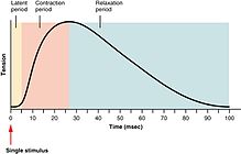
Twitch

Summation and tetanus
Three types of skeletal muscle contractions
The strength of skeletal muscle contractions can be broadly separated into twitch, summation, and tetanus. A twitch is a single wrinkle and relaxation cycle produced by an action potential within the muscle fiber itself.[26] The time between a stimulus to the motor nervus and the subsequent contraction of the innervated musculus is called the latent flow, which usually takes nearly ten ms and is acquired past the time taken for nervus action potential to propagate, the time for chemical manual at the neuromuscular junction, and then the subsequent steps in excitation-wrinkle coupling.[27]
If some other muscle activity potential were to be produced before the consummate relaxation of a muscle twitch, then the next twitch will simply sum onto the previous twitch, thereby producing a summation. Summation tin be achieved in two ways:[28] frequency summation and multiple cobweb summation. In frequency summation, the forcefulness exerted by the skeletal muscle is controlled past varying the frequency at which activeness potentials are sent to muscle fibers. Activeness potentials practice not arrive at muscles synchronously, and, during a wrinkle, some fraction of the fibers in the muscle will be firing at whatsoever given time. In a typical circumstance, when humans are exerting their muscles as hard as they are consciously able, roughly one-third of the fibers in each of those muscles will fire at once[ commendation needed ], though this ratio tin can be afflicted past diverse physiological and psychological factors (including Golgi tendon organs and Renshaw cells). This 'low' level of wrinkle is a protective machinery to forbid avulsion of the tendon—the force generated by a 95% contraction of all fibers is sufficient to harm the trunk. In multiple fiber summation, if the central nervous system sends a weak signal to contract a muscle, the smaller motor units, being more excitable than the larger ones, are stimulated first. As the strength of the signal increases, more motor units are excited in addition to larger ones, with the largest motor units having as much as 50 times the contractile forcefulness as the smaller ones. Equally more than and larger motor units are activated, the force of muscle contraction becomes progressively stronger. A concept known as the size principle, allows for a gradation of muscle force during weak contraction to occur in small steps, which so become progressively larger when greater amounts of force are required.
Finally, if the frequency of muscle activity potentials increases such that the muscle wrinkle reaches its peak force and plateaus at this level, and then the wrinkle is a tetanus.
Length-tension human relationship [edit]

Muscle length versus isometric force
Length-tension relationship relates the forcefulness of an isometric wrinkle to the length of the muscle at which the contraction occurs. Muscles operate with greatest active tension when close to an platonic length (often their resting length). When stretched or shortened beyond this (whether due to the activeness of the muscle itself or by an outside force), the maximum active tension generated decreases.[29] This decrease is minimal for pocket-sized deviations, but the tension drops off chop-chop as the length deviates further from the platonic. Due to the presence of elastic proteins inside a muscle cell (such as titin) and extracellular matrix, as the muscle is stretched beyond a given length, there is an entirely passive tension, which opposes lengthening. Combined, there is a stiff resistance to lengthening an active muscle far across the peak of active tension.
Force-velocity relationships [edit]
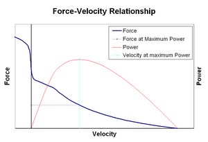
Force–velocity relationship: right of the vertical axis concentric contractions (the muscle is shortening), left of the axis eccentric contractions (the musculus is lengthened under load); power developed by the muscle in ruby. Since ability is equal to force times velocity, the muscle generates no ability at either isometric force (due to zero velocity) or maximal velocity (due to zero forcefulness). The optimal shortening velocity for ability generation is approximately one-third of maximum shortening velocity.
Force–velocity relationship relates the speed at which a muscle changes its length (usually regulated by external forces, such as load or other muscles) to the amount of force that it generates. Force declines in a hyperbolic fashion relative to the isometric force as the shortening velocity increases, eventually reaching zero at some maximum velocity. The opposite holds truthful for when the muscle is stretched – forcefulness increases to a higher place isometric maximum, until finally reaching an accented maximum. This intrinsic property of active muscle tissue plays a function in the active damping of joints that are actuated by simultaneously-active opposing muscles. In such cases, the forcefulness-velocity profile enhances the force produced by the lengthening muscle at the expense of the shortening musculus. This favoring of whichever muscle returns the joint to equilibrium effectively increases the damping of the joint. Moreover, the force of the damping increases with muscle strength. The motor arrangement can thus actively control articulation damping via the simultaneous contraction (co-contraction) of opposing musculus groups.[30]
Smoothen muscle [edit]
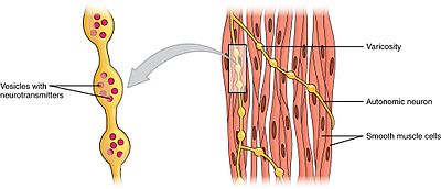
Swellings called varicosities belonging to an autonomic neuron innervate the smoothen muscle cells.
Smooth muscles can be divided into two subgroups: single-unit and multiunit. Single-unit smooth muscle cells can be found in the gut and blood vessels. Because these cells are linked together by gap junctions, they are able to contract every bit a functional syncytium. Unmarried-unit smooth muscle cells contract myogenically, which can be modulated by the autonomic nervous system.
Unlike single-unit smooth muscle cells, multiunit smooth muscle cells are constitute in the muscle of the middle and in the base of hair follicles. Multiunit smooth muscle cells contract by being separately stimulated past nerves of the autonomic nervous organisation. As such, they allow for fine command and gradual responses, much like motor unit recruitment in skeletal muscle.
Mechanisms of smoothen muscle contraction [edit]

Shine muscle contractions

Sliding filaments in contracted and uncontracted states
The contractile action of smooth muscle cells tin be tonic (sustained) or phasic (transient)[31] and is influenced by multiple inputs such as spontaneous electrical activeness, neural and hormonal inputs, local changes in chemic limerick, and stretch.[one] This is in contrast to the contractile activeness of skeletal musculus cells, which relies on a single neural input. Some types of shine muscle cells are able to generate their own action potentials spontaneously, which normally occur following a pacemaker potential or a deadening wave potential. These action potentials are generated past the influx of extracellular Ca 2+
, and not Na +
. Like skeletal muscles, cytosolic Ca ii+
ions are also required for crossbridge cycling in smooth muscle cells.
The two sources for cytosolic Ca 2+
in smooth muscle cells are the extracellular Ca ii+
entering through calcium channels and the Ca 2+
ions that are released from the sarcoplasmic reticulum. The elevation of cytosolic Ca 2+
results in more Ca 2+
bounden to calmodulin, which and so binds and activates myosin light-concatenation kinase. The calcium-calmodulin-myosin light-chain kinase complex phosphorylates myosin on the 20 kilodalton (kDa) myosin lite chains on amino acrid residue-serine 19, initiating contraction and activating the myosin ATPase. Unlike skeletal muscle cells, smooth muscle cells lack troponin, fifty-fifty though they contain the thin filament protein tropomyosin and other notable proteins – caldesmon and calponin. Thus, smooth muscle contractions are initiated by the Ca 2+
-activated phosphorylation of myosin rather than Ca 2+
bounden to the troponin complex that regulates myosin bounden sites on actin like in skeletal and cardiac muscles.
Termination of crossbridge cycling (and leaving the muscle in latch-land) occurs when myosin lite chain phosphatase removes the phosphate groups from the myosin heads. Phosphorylation of the 20 kDa myosin light chains correlates well with the shortening velocity of smoothen muscle. During this catamenia, there is a rapid burst of energy utilization equally measured past oxygen consumption. Inside a few minutes of initiation, the calcium level markedly decreases, the twenty kDa myosin light bondage' phosphorylation decreases, and free energy utilization decreases; notwithstanding, force in tonic smooth muscle is maintained. During contraction of muscle, rapidly cycling crossbridges form between activated actin and phosphorylated myosin, generating forcefulness. It is hypothesized that the maintenance of force results from dephosphorylated "latch-bridges" that slowly cycle and maintain force. A number of kinases such as rho kinase, DAPK3, and protein kinase C are believed to participate in the sustained phase of contraction, and Ca 2+
flux may be significant.
Neuromodulation [edit]
Although smooth muscle contractions are myogenic, the rate and strength of their contractions can be modulated by the autonomic nervous system. Postganglionic nerve fibers of parasympathetic nervous system release the neurotransmitter acetylcholine, which binds to muscarinic acetylcholine receptors (mAChRs) on smooth muscle cells. These receptors are metabotropic, or G-poly peptide coupled receptors that initiate a second messenger cascade. Conversely, postganglionic nerve fibers of the sympathetic nervous organisation release the neurotransmitters epinephrine and norepinephrine, which bind to adrenergic receptors that are also metabotropic. The verbal effects on the smooth musculus depend on the specific characteristics of the receptor activated—both parasympathetic input and sympathetic input can be either excitatory (contractile) or inhibitory (relaxing).
Cardiac muscle [edit]
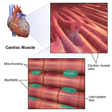
At that place are two types of cardiac muscle cells: autorhythmic and contractile. Autorhythmic cells do non contract, but instead set the footstep of wrinkle for other cardiac muscle cells, which can be modulated past the autonomic nervous system. In dissimilarity, contractile muscle cells (cardiomyocytes) institute the bulk of the heart muscle and are able to contract.
Excitation-contraction coupling [edit]
In both skeletal and cardiac muscle excitation-contraction (E-C) coupling, depolarization conduction and Ca2+ release processes occur. However, though the proteins involved are like, they are distinct in structure and regulation. The dihydropyridine receptors (DHPRs) are encoded by different genes, and the ryanodine receptors (RyRs) are distinct isoforms. Besides, DHPR contacts with RyR1 (main RyR isoform in skeletal musculus) to regulate Ca2+ release in skeletal musculus, while the L-blazon calcium channel (DHPR on cardiac myocytes) and RyR2 (main RyR isoform in cardiac muscle) are non physically coupled in cardiac muscle, merely face with each other by a junctional coupling.[32]
Dissimilar skeletal muscle, E-C coupling in cardiac musculus is thought to depend primarily on a machinery called calcium-induced calcium release,[33] which is based on the junctional construction betwixt T-tubule and sarcoplasmic reticulum. Junctophilin-2 (JPH2) is essential to maintain this construction, besides as the integrity of T-tubule.[34] [35] [36] Another protein, receptor accessory protein 5 (REEP5), functions to keep the normal morphology of junctional SR.[37] Defects of junctional coupling can result from deficiencies of either of the two proteins. During the process of calcium-induced calcium release, RyR2s are activated by a calcium trigger, which is brought almost by the flow of Caii+ through the Fifty-type calcium channels. Later on this, cardiac muscle tends to exhibit diad structures, rather than triads.
Excitation-contraction coupling in cardiac muscle cells occurs when an action potential is initiated past pacemaker cells in the sinoatrial node or Atrioventricular node and conducted to all cells in the center via gap junctions. The activeness potential travels along the surface membrane into T-tubules (the latter are not seen in all cardiac cell types) and the depolarisation causes extracellular Ca two+
to enter the prison cell via Fifty-type calcium channels and possibly sodium-calcium exchanger (NCX) during the early part of the plateau phase. Although this Ca2+ influx only count for about 10% of the Catwo+ needed for activation, it is relatively larger than that of skeletal musculus. This Ca 2+
influx causes a small local increase in intracellular Ca ii+
. The increment of intracellular Ca two+
is detected by RyR2 in the membrane of the sarcoplasmic reticulum, which releases Ca 2+
in a positive feedback physiological response. This positive feedback is known as calcium-induced calcium release[33] and gives rise to calcium sparks (Ca 2+
sparks[38]). The spatial and temporal summation of ~30,000 Ca two+
sparks gives a cell-wide increase in cytoplasmic calcium concentration.[39] The increase in cytosolic calcium following the flow of calcium through the cell membrane and sarcoplasmic reticulum is moderated past calcium buffers, which bind a large proportion of intracellular calcium. As a effect, a large increase in total calcium leads to a relatively small rise in free Ca 2+
.[twoscore]
The cytoplasmic calcium binds to Troponin C, moving the tropomyosin circuitous off the actin binding site allowing the myosin head to bind to the actin filament. From this point on, the contractile mechanism is essentially the same every bit for skeletal muscle (higher up). Briefly, using ATP hydrolysis, the myosin head pulls the actin filament toward the center of the sarcomere.

Fundamental proteins involved in cardiac calcium cycling and excitation-contraction coupling
Post-obit systole, intracellular calcium is taken up by the sarco/endoplasmic reticulum ATPase (SERCA) pump back into the sarcoplasmic reticulum ready for the next cycle to brainstorm. Calcium is besides ejected from the cell mainly by the sodium-calcium exchanger (NCX) and, to a lesser extent, a plasma membrane calcium ATPase. Some calcium is also taken up by the mitochondria.[41] An enzyme, phospholamban, serves as a brake for SERCA. At depression heart rates, phospholamban is active and slows down the activity of the ATPase so that Ca two+
does not have to leave the prison cell entirely. At loftier heart rates, phospholamban is phosphorylated and deactivated thus taking most Ca two+
from the cytoplasm back into the sarcoplasmic reticulum. In one case again, calcium buffers moderate this fall in Ca 2+
concentration, permitting a relatively minor subtract in free Ca 2+
concentration in response to a big change in total calcium. The falling Ca 2+
concentration allows the troponin complex to dissociate from the actin filament thereby catastrophe contraction. The heart relaxes, assuasive the ventricles to fill with blood and begin the cardiac bike again.
Invertebrates [edit]
Round and longitudinal muscles [edit]

A simplified image showing earthworm movement via peristalsis
In annelids such as earthworms and leeches, circular and longitudinal muscles cells grade the body wall of these animals and are responsible for their movement.[42] In an earthworm that is moving through a soil, for case, contractions of circular and longitudinal muscles occur reciprocally while the coelomic fluid serves as a hydroskeleton past maintaining turgidity of the earthworm.[43] When the circular muscles in the anterior segments contract, the inductive portion of fauna's body begins to constrict radially, which pushes the incompressible coelomic fluid forrard and increasing the length of the fauna. As a result, the front end of the beast moves forward. As the forepart of the earthworm becomes anchored and the round muscles in the anterior segments become relaxed, a wave of longitudinal muscle contractions passes backwards, which pulls the rest of animal'south abaft body forrad.[42] [43] These alternate waves of circular and longitudinal contractions is called peristalsis, which underlies the creeping movement of earthworms.
Obliquely striated muscles [edit]
Invertebrates such as annelids, mollusks, and nematodes, possess obliquely striated muscles, which comprise bands of thick and sparse filaments that are bundled helically rather than transversely, like in vertebrate skeletal or cardiac muscles.[44] In bivalves, the obliquely striated muscles can maintain tension over long periods without using too much energy. Bivalves use these muscles to keep their shells closed.
Asynchronous muscles [edit]
Asynchronous muscles power flying in most insect species. a: Wings b: Wing articulation c: Dorsoventral muscles power the upstroke d: Dorsolongitudinal muscles (DLM) power the downstroke. The DLMs are oriented out of the page.
Advanced insects such as wasps, flies, bees, and beetles possess asynchronous muscles that constitute the flight muscles in these animals.[44] These flight muscles are often called fibrillar muscles because they contain myofibrils that are thick and conspicuous.[45] A remarkable feature of these muscles is that they do not crave stimulation for each muscle contraction. Hence, they are called asynchronous muscles considering the number of contractions in these muscles exercise non correspond (or synchronize) with the number of action potentials. For example, a wing muscle of a tethered fly may receive action potentials at a frequency of three Hz but it is able to beat at a frequency of 120 Hz.[44] The loftier frequency beating is made possible because the muscles are continued to a resonant system, which is driven to a natural frequency of vibration.
History [edit]

Electrodes touch on a frog, and the legs twitch into the upward position[46]
In 1780, Luigi Galvani discovered that the muscles of dead frogs' legs twitched when struck by an electrical spark.[47] This was i of the first forays into the written report of bioelectricity, a field that nonetheless studies the electrical patterns and signals in tissues such as fretfulness and muscles.
In 1952, the term excitation–contraction coupling was coined to draw the physiological process of converting an electrical stimulus to a mechanical response.[xx] This procedure is primal to musculus physiology, whereby the electric stimulus is ordinarily an action potential and the mechanical response is contraction. Excitation–contraction coupling can be dysregulated in many diseases. Though excitation–wrinkle coupling has been known for over half a century, it is all the same an active surface area of biomedical research. The general scheme is that an action potential arrives to depolarize the cell membrane. By mechanisms specific to the musculus blazon, this depolarization results in an increase in cytosolic calcium that is called a calcium transient. This increase in calcium activates calcium-sensitive contractile proteins that and so use ATP to cause cell shortening.
The mechanism for muscle contraction evaded scientists for years and requires continued research and updating.[48] The sliding filament theory was independently developed past Andrew F. Huxley and Rolf Niedergerke and by Hugh Huxley and Jean Hanson. Their findings were published equally ii consecutive papers published in the 22 May 1954 issue of Nature under the common theme "Structural Changes in Muscle During Contraction".[22] [23]
See also [edit]
- Anatomical terms of motion
- calcium-induced calcium release
- Cardiac activeness potential
- Cramp
- Dystonia
- Practice physiology
- Fasciculation
- Loma's muscle model
- Hypnic jerk
- In vitro muscle testing
- Lombard'due south paradox
- Myoclonus
- Rigor mortis
- Spasm
- Uterine contraction
References [edit]
- ^ a b c d e f chiliad h i j k l m n o Widmaier, Eric P.; Raff, Hersel; Strang, Kevin T. (2010). "Muscle". Vander'south Homo Physiology: The Mechanisms of Body Function (twelfth ed.). New York, NY: McGraw-Loma. pp. 250–291. ISBN978-0-321-98122-half-dozen.
- ^ Silverthorn, Dee Unglaub (2016). "Muscles". Man Physiology: An Integrated Arroyo (seventh ed.). San Francisco, CA: Pearson. pp. 377–416. ISBN978-0-321-98122-6.
- ^ a b c d e f Aidley, David J. (1998). "Mechanics and energetics of muscular contraction". The Physiology of Excitable Cells (quaternary ed.). New York, NY: Cambridge Academy Printing. pp. 323–335. ISBN978-0-521-57421-1.
- ^ a b c d eastward f Sircar, Sabyasachi (2008). "Muscle elasticity". Principles of Medical Physiology (1st ed.). New York, NY: Thieme. p. 113. ISBN978-1-588-90572-seven.
- ^ a b c d due east f Bullock, John; Boyle, Joseph; Wang, Michael B. (2001). "Muscle contraction". NMS Physiology. Vol. 578 (4th ed.). Baltimore, Maryland: Lippincott Williams and Wilkins. pp. 37–56.
- ^ a b Kumar, Shrawan (2008). "Introduction and terminology". In Shrawan Kumar (ed.). Muscle forcefulness (1st ed.). Boca Raton, FL: CRC Press. p. 113. ISBN978-0-415-36953-4.
- ^ a b Biewener, Andrew A. (2003). "Muscles and skeletons: The building blocks of animal movement". Animate being Locomotion. Oxford Fauna Biological science Series. New York, NY: Oxford Academy Printing. pp. 15–45. ISBN978-0-198-50022-three.
- ^ Faulkner JA (2003). "Terminology for contractions of muscles during shortening, while isometric, and during lengthening". Journal of Applied Physiology. 95 (2): 455–459. doi:10.1152/japplphysiol.00280.2003. PMID 12851415.
- ^ a b "Types of contractions". 2006-05-31. Retrieved 2007-10-02 .
- ^ a b c Colliander EB, Tesch PA (1990). "Effects of eccentric and concentric muscle deportment in resistance training". Acta Physiol. Scand. 140 (ane): 31–9. doi:x.1111/j.1748-1716.1990.tb08973.x. PMID 2275403.
- ^ Nikolaidis MG, Kyparos A, Spanou C, Paschalis V, Theodorou AA, Vrabas IS (2012). "Redox biology of exercise: an integrative and comparative consideration of some overlooked issues". J. Exp. Biol. 215 (Pt ten): 1615–25. doi:x.1242/jeb.067470. PMID 22539728.
- ^ Schmidt-Rohr, One thousand. (2020). "Oxygen Is the High-Free energy Molecule Powering Complex Multicellular Life: Primal Corrections to Traditional Bioenergetics ACS Omega 5: 2221-2233. http://dx.doi.org/10.1021/acsomega.9b03352
- ^ Brooks, Thou.A; Fahey, T.D.; White, T.P. (1996). Exercise Physiology: Human Bioenergetics and Its Applications. (second ed.). Mayfield Publishing Co.
- ^ Alfredson, H; Pietilä, T; Jonsson, P; Lorentzon, R (1998). "Heavy-load eccentric dogie muscle training for the treatment of chronic Achilles tendinosis" (PDF). The American Journal of Sports Medicine. 26 (3): 360–6. doi:ten.1177/03635465980260030301. PMID 9617396. S2CID 30259362.
- ^ Satyendra L, Byl N (2006). "Effectiveness of physical therapy for Achilles tendinopathy: An show based review of eccentric exercises". Isokinetics and Exercise Science. 14 (1): 71–80. doi:10.3233/IES-2006-0223.
- ^ Cannell LJ, Taunton JE, Clement DB, Smith C, Khan KM (2001). "A randomised clinical trial of the efficacy of drop squats or leg extension/leg ringlet exercises to treat clinically diagnosed jumper'due south knee in athletes: pilot study". Br J Sports Med. 35 (ane): 60–iv. doi:10.1136/bjsm.35.1.lx. PMC1724276. PMID 11157465.
- ^ Tassinary; Cacioppo (2000). "The Skeletomotor organisation: surface electromyography". In Cacioppo, John T.; Tassinary, Luois 1000.; Berntson, Gary G. (eds.). Handbook of Psychophysiology (Second ed.). Cambridge: Cambridge University Press. ISBN978-0-521-62634-seven.
- ^ Levitan, Irwin; Kaczmarek, Leonard (August 19, 2015). "Intercellular communication". The Neuron: Cell and Molecular Biology (4th ed.). New York, NY: Oxford Academy Press. pp. 153–328. ISBN978-0199773893.
- ^ a b Saladin, Kenneth S., Stephen J. Sullivan, and Christina A. Gan. Beefcake & Physiology: The Unity of Grade and Part. 7th ed. New York: McGraw-Hill Education, 2015. Print.
- ^ a b Sandow A (1952). "Excitation-Contraction Coupling in Muscular Response". Yale J Biol Med. 25 (3): 176–201. PMC2599245. PMID 13015950.
- ^ Saladin, Kenneth (2012). Beefcake and Physiology: The Unity of Form and Function. New York: McGraw Hill. ISBN978-0-07-337825-1.
- ^ a b Huxley AF, Niedergerke R (1954). "Structural Changes in Musculus During Contraction: Interference Microscopy of Living Muscle Fibres". Nature. 173 (4412): 971–973. Bibcode:1954Natur.173..971H. doi:10.1038/173971a0. PMID 13165697. S2CID 4275495.
- ^ a b Huxley H, Hanson J (1954). "Changes in the cross-striations of musculus during contraction and stretch and their structural interpretation". Nature. 173 (4412): 973–976. Bibcode:1954Natur.173..973H. doi:10.1038/173973a0. PMID 13165698. S2CID 4180166.
- ^ Horowits R, Podolsky RJ (November 1987). "The positional stability of thick filaments in activated skeletal musculus depends on sarcomere length: prove for the part of titin filaments". J. Jail cell Biol. 105 (5): 2217–23. doi:10.1083/jcb.105.v.2217. PMC2114850. PMID 3680378.
- ^ a b c Enoka, Roger M.; Pearson, Keir G. (2013). "The motor unit and muscle action". In Eric R. Kandel; James H. Schwartz; Thomas Thousand. Jessell; Steven A. Siegelbaum; A. J. Hudspeth (eds.). Principles of Neural Scientific discipline (5th ed.). New York, NY: McGraw-Hill Medical. pp. 768–789. ISBN978-0-071-39011-8.
- ^ Feher, Joseph (2012). "Chapter iii.4: Skeletal muscle mechanics". Quantitative Human Physiology: An Introduction. Bookish Press Serial in Biomedical Engineering (1st ed.). New York, NY: Bookish Press. pp. 239–248. ISBN978-0-123-82163-viii.
- ^ Khurana, Indu (2006). "Characteristics of muscle excitability and contractility". Textbook Of Medical Physiology (1st ed.). Elsevier. pp. 101–2.
- ^ Shwedyk, E.; Balasubramanian, R.; Scott, R. Due north. (1977). "A nonstationary model for the Electromyogram". IEEE Transactions on Biomedical Engineering. 24 (5): 417–424. doi:10.1109/TBME.1977.326175. PMID 892834. S2CID 1770255.
- ^ Gordon AM, Huxley AF, Julian FJ (1966). "The variation in isometric tension with sarcomere length in vertebrate muscle fibres". J. Physiol. 184 (1): 170–92. doi:10.1113/jphysiol.1966.sp007909. PMC1357553. PMID 5921536.
- ^ Heitmann, Stewart; Ferns, Norm; Breakpsear, Michael (2011). "Muscle co-contraction modulates damping and joint stability in a iii-link biomechanical limb". Frontiers in Neurorobotics. 5: 5. doi:x.3389/fnbot.2011.00005. ISSN 1662-5218. PMC3257849. PMID 22275897.
- ^ Zhang, Y; Hermanson, ME; Eddinger, TJ (2013). "Tonic and phasic smooth musculus contraction is non regulated by the PKCα - CPI-17 pathway in swine breadbasket antrum and fundus". PLOS ONE. viii (9): e74608. Bibcode:2013PLoSO...874608Z. doi:x.1371/journal.pone.0074608. PMC3776813. PMID 24058600.
- ^ Martonosi, Anthony N.; Pikula, Slawomir (2003). "The network of calcium regulation in muscle". Acta Biochimica Polonica. l (1): one–30. doi:10.18388/abp.2003_3711. ISSN 0001-527X. PMID 12673344.
- ^ a b Fabiato, A. (1983). "Calcium-induced calcium release from the cardiac sarcoplasmic reticulum". American Journal of Physiology. 245 (i): C1–14. doi:10.1152/ajpcell.1983.245.1.C1. PMID 6346892.
- ^ Guo, Ang; Zhang, Xiaoying; Iyer, Venkat Ramesh; Chen, Biyi; Zhang, Caimei; Kutschke, William J.; Weiss, Robert Yard.; Franzini-Armstrong, Clara; Song, Long-Sheng (2014-08-xix). "Overexpression of junctophilin-ii does non enhance baseline function but attenuates heart failure development later cardiac stress". Proceedings of the National Academy of Sciences of the United States of America. 111 (33): 12240–12245. Bibcode:2014PNAS..11112240G. doi:10.1073/pnas.1412729111. ISSN 1091-6490. PMC4143026. PMID 25092313.
- ^ Wei, Sheng; Guo, Ang; Chen, Biyi; Kutschke, William; Xie, Yu-Ping; Zimmerman, Kathy; Weiss, Robert M.; Anderson, Mark Due east.; Cheng, Heping; Song, Long-Sheng (2010-08-xx). "T-tubule remodeling during transition from hypertrophy to eye failure". Apportionment Research. 107 (iv): 520–531. doi:10.1161/CIRCRESAHA.109.212324. ISSN 1524-4571. PMC2927862. PMID 20576937.
- ^ Takeshima, H.; Komazaki, S.; Nishi, M.; Iino, M.; Kangawa, K. (July 2000). "Junctophilins: a novel family of junctional membrane complex proteins". Molecular Cell. 6 (1): xi–22. doi:ten.1016/s1097-2765(00)00003-4. ISSN 1097-2765. PMID 10949023.
- ^ Yao, Lei; Xie, Duanyang; Geng, Li; Shi, Dan; Huang, Jian; Wu, Yufei; Lv, Fei; Liang, Dandan; Li, Li; Liu, Yi; Li, Jun (3 February 2018). "REEP5 (Receptor Accompaniment Protein 5) Acts as a Sarcoplasmic Reticulum Membrane Sculptor to Modulate Cardiac Function". Periodical of the American Heart Association. 7 (3). doi:10.1161/JAHA.117.007205. ISSN 2047-9980. PMC5850239. PMID 29431104.
- ^ Cheng H, Lederer WJ, Cannell MB (October 1993). "Calcium sparks: simple events underlying excitation-contraction coupling in heart muscle". Science. 262 (5134): 740–4. Bibcode:1993Sci...262..740C. doi:10.1126/scientific discipline.8235594. PMID 8235594.
- ^ Cannell MB, Cheng H, Lederer WJ (November 1994). "Spatial not-uniformities in Ca 2+
i during excitation-contraction coupling in cardiac myocytes". Biophys. J. 67 (five): 1942–56. Bibcode:1994BpJ....67.1942C. doi:10.1016/S0006-3495(94)80677-0. PMC1225569. PMID 7858131. - ^ Thousand., Bers, D. (2001). Excitation-contraction coupling and cardiac contractile force (2nd ed.). Dordrecht: Kluwer Bookish Publishers. ISBN9780792371571. OCLC 47659382.
- ^ Crespo LM, Grantham CJ, Cannell MB (June 1990). "Kinetics, stoichiometry and role of the Na-Ca commutation mechanism in isolated cardiac myocytes". Nature. 345 (6276): 618–21. Bibcode:1990Natur.345..618C. doi:10.1038/345618a0. PMID 2348872. S2CID 4348240.
- ^ a b Hillis, David Thou.; Sadava, David E.; Price, Mary Five. (2014). "Muscle and movement". Principles of Life (2nd ed.). Sunderland, MA: Sinauer Associates. pp. 681–698. ISBN978-i-464-10947-8.
- ^ a b Gardner, C.R. (1976). "The neuronal control of locomotion in the earthworm". Biological Reviews of the Cambridge Philosophical Guild. 51 (1): 25–52. doi:ten.1111/j.1469-185X.1976.tb01119.x. PMID 766843. S2CID 9983649.
- ^ a b c Alexander, R. McNeill (2003). "Muscle, the motor". Principles of Beast Locomotion (2nd ed.). Princeton, NJ: Princeton University Press. pp. 15–37. ISBN978-0-691-12634-0.
- ^ Josephson, R. K.; Malamud, J. G.; Stokes, D. R. (2000-09-15). "Asynchronous muscle: a primer". Periodical of Experimental Biological science. 203 (eighteen): 2713–2722. doi:10.1242/jeb.203.18.2713. ISSN 0022-0949. PMID 10952872.
- ^ David Ames Wells, The science of common things: a familiar explanation of the first, 323 pages (page 290)
- ^ Whittaker, E. T. (1951), A History of the Theories of Aether and Electricity. Vol 1, Nelson, London
- ^ Huxley, H. E. (April 2000). "Past, Present and Future Experiments on Muscle". Philosophical Transactions: Biological Sciences. 355 (1396): 539–543. doi:10.1098/rstb.2000.0595. JSTOR 3066716. PMC1692762. PMID 10836507.
Further reading [edit]
- Saladin, Kenneth S., Stephen J. Sullivan, and Christina A. Gan. (2015). Anatomy & Physiology: The Unity of Form and Function. seventh ed. New York: McGraw-Hill Educational activity.
- Krans, J. 50. (2010) The Sliding Filament Theory of Muscle Contraction. Nature Education three(ix):66
External links [edit]
- Sliding Filament Model of Muscle Wrinkle
- Animation: Myofilament Contraction

Source: https://en.wikipedia.org/wiki/Muscle_contraction
Posted by: steffeylooncomet.blogspot.com
0 Response to "What Type Of Contraction Involves The Development Of Tension But No Change In Length"
Post a Comment